Departments
Our Radiologists
Department of Neuroradiology
The Department of Neuroradiology at Citi Neuro Centre is committed to provide state of the art diagnostic and interventional radiological services. Our experienced technicians and neuroradiologists are equipped with the right training and skills to provide prompt and accurate diagnosis. As a department that lends crucial support to all other specialities, we believe in working as a team to provide high-quality, patient-centric diagnostic services to patients.
Interventional Radiology
Apart from diagnostic services, the department offers full-spectrum of neurovascular, peripheral vascular and non-vascular interventional procedures including management of brain aneurysms, vascular malformations and several other neurological conditions, Peripheral angioplasty and stenting of blood vessels (Peripheral, Carotid, Renal, Visceral), Tumour Embolisations including Chemo Embolisations, Fibroid Embolisations, etc. It also works closely with the integrated Pain Management program that includes image guided nerve root and facet blocks and percutaneous vertebroplasties.
Technology
The department is equipped with:
- 3 Tesla, Fully Digital MRI Scanner - A fully digital high-field MRI system with high performance gradient systems (amongst the most powerful and advanced units in the world). The unit provides exceptional image quality with shorter scan duration. The wide bore of the MRI unit as well as the interiors of the MRI room have been modelled to reduce patient anxiety and claustrophobia. The system has advanced imaging protocols to facilitate accurate diagnosis in neuro, cardiac, musculoskeletal and body imaging.
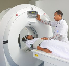
The system, which is has several cutting edge technologies such as - Ultrafast imaging protocols, Motion correction techniques that allow imaging of irritable and uncooperative patients, brain diffusion and perfusion studies, Arterial spin labelling, Functional MRI, MR spectroscopy, Diffusion tensor imaging, Whole body diffusion studies, Advanced cardiac imaging applications include myocardial perfusion and viability studies and Non-contrast MR Angiography - Multidetector CT scanner - Ensures maximum acquisition efficiency, providing speed, resolution, image quality and extended coverage with minimum possible radiation exposure. It has optimized application protocols for brain and spine imaging in uncooperative patients, high resolution 3D imaging of small structures, CT scan of joints, CT Angiography, perfusion imaging, and CT guided interventional procedures.
- Colour Doppler Units - Most sophisticated Colour Doppler Systems with lightweight probes for Radiology, Vascular, Neuro and Cardiac applications. The system gives high resolution images in any plane to reveal the smallest details with stunning clarity.
- Computed Radiography - Renders high-resolution radiographs with superior time efficiency. As no repetition and digital enhancement are required, rapid transfer of images for immediate investigation by physician is made possible through integration with PACS.
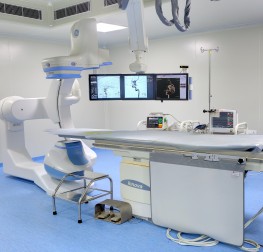
- Advanced neurointerventional suite with 3D rotational angiography - Enables digital subtraction angiography which permits clear visualization of brain, spine and coronary blood vessels without interfence from bony or dense soft tissue environment.
3D rotational angiography provides an image of a particular vessel while the camera rotates around the patient in a predefined arc, enabling viewing of the structure from multiple angles with a single injection of contrast medium. It gives exquisitely detailed 3 Dimensional visualization of the blood vessels, thereby enabling faster and more accurate clinical decisions while treating complex abnormalities of the blood vessels. - Picture Archival and Communications System (PACS) - There is a local network connecting all imaging modalities like CR, CT, MRI and Cath lab to a common workstation. This facilitates faster transfer of the images to the radiologist's and clinician's desktop computer. This allows faster and more efficient reporting, faster clinical decisions and has drastically changed the way patients receive healthcare.
Investigations and Procedures Performed
- Whole Body CT Scan
- CT Angiogram
- CT Perfusion
- CT Guided Biopsy
- CT Guided Pain Procedures (Nerve / Facet blocks)
- CT Cisternogram
- CT Myelogram
- Whole Body MRI
- Whole Body Diffusion MRI
- Perfusion Imaging including ASL
- Functional MRI
- Epilepsy Imaging
- MR Spectroscopy
- MR Neurography
- Peripheral Angioplasty and Stenting
- Renal artery stenting and angioplasty
- Vascular malformations - Embolization
- Vascular malformations - Sclerotherapy
- Preoperative embolization of Tumors
- Transarterial chemoembolization
- Vertebroplasty / Cementoplasty
- Bronchial artery Embolisation
- Embolization for epistaxis
- Embolization for Intestinal Bleeding
- Embolization for Uterine bleeding
- Percutaneous Cholecystostomy
- Biliary Drainage and Stenting
- Interventions for Liver Cancer
- Transarterial chemoembolization
- Mechanical thrombectomy for Arterial Ischemic Stroke
- Intraarterial Infusion Therapy for Vasospasm
- Carotid angioplasty and stenting
- Subclavian - Vertebral angioplasty and stenting
- Intracranial angioplasty and stenting
- Coil Embolization for Brain Aneurysms
- Balloon Assisted Coil Embolization for Brain Aneurysms
- Stent Assisted Coil Embolization for Brain Aneurysms
- Flow Diverter Placement for Brain Aneurysms
- Parent artery occlusion
- Embolization for Brain AV Malformations
- Embolization for Carotid Cavernous Fistula
- Embolization for Dural AV Fistula
- Dural sinus thrombolysis for Venous Sinus Thrombosis
- Embolization for Brain Tumors
- Embolization for Spinal Vascular Malformations
- Embolization for Spinal Tumors
- Percutaneous Vertebroplasty and Sacroplasty
- Pain Management Procedures
- Embolization for for Nasal Bleeds
- Embolization for Paragangliomas
- Embolization for for Neck Tumors
- Embolization for Arteriovenous malformations of face and scalp